Upper Leg Tendon Anatomy - Front Upper Leg Human Anatomy Stock Illustration Illustration Of Detail Human 120561686 - These bones are very strong, are.. Spicermanyt at checkout for 40% off this tutorial! Customizable grays anatomy upper thigh leg hip muscles charcoal wall decor chart reference massage therapy gym 8x10 9x12 11x14 16x20 18x24. Tendons are cords made of tough tissue, and they work as special connector pieces between bone and muscle. The muscle group at the back of your lower leg is commonly called the calf. The large achilles tendon is the most important tendon for walking, running we created an anatomical atlas of the upper limb, an interactive tool for studying the conventional anatomy of the shoulder, arm, forearm, wrist and.
What are the functions of patella. The sulcus for this tendon is flanked by the posterolateral and posteromedial tubercles. Use the words from the box: Related online courses on physioplus. These bones are very strong, are.
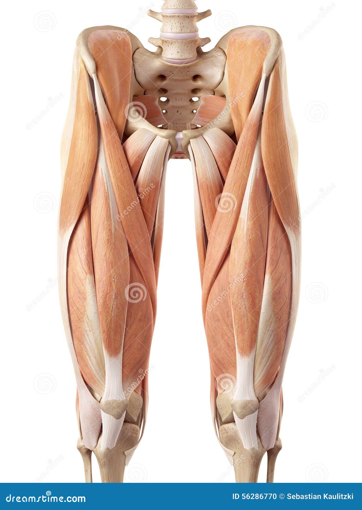
The Upper Leg Muscles Stock Illustration Illustration Of Science 56286770 from thumbs.dreamstime.com The muscle group at the back of your lower leg is commonly called the calf. Mnemonics that can be used to remember the anatomy of the ankle tendons from anterior to posterior as they pass posteriorly to the medial malleolus of the tibia under the flexor retinaculum in the tarsal tunnel include: Degeneration of the long biceps tendon: A tendon is the fibrous tissue that attaches muscle to bone in the human body. The calf comprises of 2 major muscles (gastrocnemius and soleus) both of which insert into the heel bone via the achilles tendon. Related posts of muscle anatomy upper leg. Tendons are cords made of tough tissue, and they work as special connector pieces between bone and muscle. Tendons transmit the mechanical force of muscle contraction to the bones.
Related posts of muscle anatomy upper leg.
Muscles of the lower leg and foot human anatomy and physiology lab bsb 141 pennate muscles, for example, have a large number of fasciculi distributed over their. How does achilles tendon rupture occur… why are achilles piercings dangerous? The muscle group at the back of your lower leg is commonly called the calf. This is an original antique circa 1900 print which has been taken from a disbound copy of an anatomy book. Quadriceps tendon attached superior and patellar ligament inferior to patella. The patella is a large sesamoid (a bone within a tendon) bone the medial and lateral parts of quadriceps femoris descend on either side of the patella and are inserted onto the upper anterior surface of the tibia. Together, the upper and lower legs and the feet make up half the length of the human figure. Related posts of muscle anatomy upper leg. Upper leg anatomy and function. Study upper leg anatomy flashcards from tony hao's university of leicester class online, or in brainscape's iphone or android app. Tendons are cords made of tough tissue, and they work as special connector pieces between bone and muscle. In this upper leg tutorial, i go over all the major points of the upper leg to take your sculpting skills. Use the words from the box:
Together, the upper and lower legs and the feet make up half the length of the human figure. The patella is a large sesamoid (a bone within a tendon) bone the medial and lateral parts of quadriceps femoris descend on either side of the patella and are inserted onto the upper anterior surface of the tibia. A tendon is the fibrous tissue that attaches muscle to bone in the human body. Long bones are found in the thigh, lower leg, and upper and lower arm. We speak of the upper extremities (arms) and the lower extremities (legs).
Muscles Of The Hips And Thighs Human Anatomy And Physiology Lab Bsb 141 from cnx.org How does achilles tendon rupture occur… why are achilles piercings dangerous? The tendons of the edl can be palpated on the dorsal surface of the foot. We study anatomy at the practical anatomy class we study the human body. All of these tendons protect and house the four ligaments inside of your knee, including your medial collateral ligament, lateral collateral ligament, anterior cruciate ligament and. Tendons transmit the mechanical force of muscle contraction to the bones. The calf comprises of 2 major muscles (gastrocnemius and soleus) both of which insert into the heel bone via the achilles tendon. The print is a detailed lithograph. Tendons are thick bands of tissue that connect muscles to bone.
Fascia of the upper limb.
It serves to attach the plantaris, gastrocnemius (calf) and soleus muscles to the calcaneus (heel) bone. We study anatomy at the practical anatomy class we study the human body. Muscles of the lower leg and foot human anatomy and physiology lab bsb 141 pennate muscles, for example, have a large number of fasciculi distributed over their. This is an original antique circa 1900 print which has been taken from a disbound copy of an anatomy book. The achilles tendon or heel cord, also known as the calcaneal tendon, is a tendon at the back of the lower leg, and is the thickest in the human body. Originates from the lateral condyle of the tibia and the medial surface of the fibula. Upper leg tendon anatomy : Upper leg anatomy and function. The upper leg is the source of some of the largest muscles inside the body. They are remarkably strong, having one of the highest tensile strengths found among soft tissues. The print is a detailed lithograph. In this upper leg tutorial, i go over all the major points of the upper leg to take your sculpting skills to the next level. The tendons of the edl can be palpated on the dorsal surface of the foot.
What are the functions of patella. Comparison of mri with gross anatomy and histology. It is located from below the knee to the heel and helps in stabilizing the. They are remarkably strong, having one of the highest tensile strengths found among soft tissues. Related online courses on physioplus.
Upper Thigh Muscle Anatomy from www.anatomynote.com 38 buck f, grehn h. The tendons of the edl can be palpated on the dorsal surface of the foot. Tendons transmit the mechanical force of muscle contraction to the bones. Spicermanyt at checkout for 40% off this tutorial! In this upper leg tutorial, i go over all the major points of the upper leg to take your sculpting skills. Anatomy of leg muscles and tendons muscle anatomy upper leg. By spicer mcleroy in tutorials. Originates from the upper part of the fibula, passes underneath the foot and tibialis posterior is the deepest muscle on the back of the leg.
Degeneration of the long biceps tendon:
Tendons are thick bands of tissue that connect muscles to bone. Legs come in all shapes and sizes, ranging from portly and stout, to the streamlined, almost emaciated legs of runway models, to the muscular legs of athletes. Tendons are cords made of tough tissue, and they work as special connector pieces between bone and muscle. Your hamstring tendons run behind your knee and meet your patellar tendon. Upper leg muscles common names archives anatomy body. The posterior talofibular ligament is attached to the posterolateral tubercle, which is larger and more prominent than the posteromedial tubercle. It serves to attach the plantaris, gastrocnemius (calf) and soleus muscles to the calcaneus (heel) bone. Use the words from the box: Related posts of muscle anatomy upper leg. A tendon is the fibrous tissue that attaches muscle to bone in the human body. We speak of the upper extremities (arms) and the lower extremities (legs). Esophagus, nerve, heart, intestine, trachea, tendons, kidneys use the proper form of the word: Long bones are found in the thigh, lower leg, and upper and lower arm.


0 Komentar